Stories

- Article
The epilepsy diagnosis
Epilepsy exists between the mind and body, something that Aparna Nair experienced for herself when she was diagnosed as a teenager.
- Article
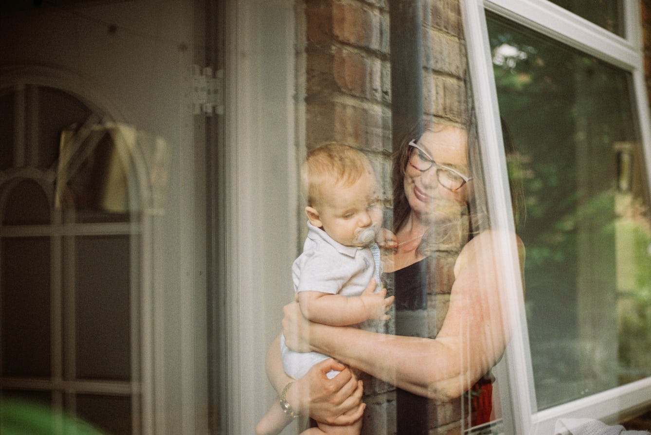
Birth, babies and boxes of memories
With memories of her baby in neonatal intensive care still fresh, Erin Beeston decides to unearth the poignant objects her family kept following births, going back as far as Victorian times.

- Article
Air of threat
Novelist Chloe Aridjis vividly describes the suffocating atmosphere of Mexico City, as a combination of topography, crowded neighbourhoods, and reckless political diktats create a downward spiral.

- Article
The food diary and the power of unhealth
Food diaries might appear to present a strictly factual record of dietary choices, but what they don’t include is the more revealing story, as Virginia Hartley suggests.


Catalogue

- Digital Images
- Online
A microCT 3D reconstruction of a 10-day-old chick embryo, as seen from the right hand side. The inner ear is depicted, with the semicircular canals (the body's balance organ) and the cochlea (which converts sound waves into electrical impulses) shown in green. The otic capsule, a cartilaginous structure surrounding the inner ear which develops into part of the sphenoid bone, is shown in blue.
Akshay Kumar, Tom Davies and Nobue Itasaki, University of Bristol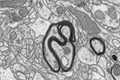
- Digital Images
- Online
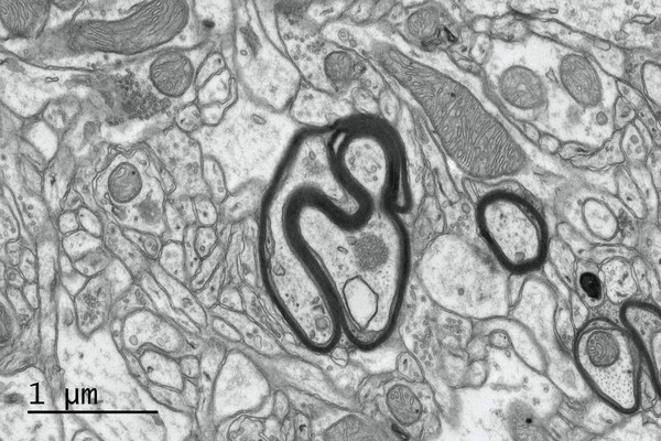
Myelinated nerves in a mouse brain, TEM
Mikaela Laine, University of Helsinki
- Digital Images
- Online
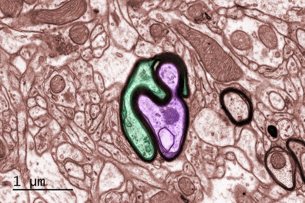
Myelinated nerves in a mouse brain, TEM
Mikaela Laine, University of Helsinki
- Digital Images
- Online
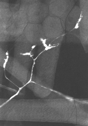
Muscle Innervation in Drosophila
Hermann Aberle, University of Munster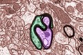
- Digital Images
- Online
Myelinated nerves in a mouse brain, TEM
Mikaela Laine, University of Helsinki